 Located in the SAVS Facility
GLAUCOMA
The term glaucoma refers to an elevated intraocular pressure inside the eye that is associated with degeneration of the retina and optic nerve. This degeneration eventually leads to blindness.
In a normal eye, fluid is constantly produced behind the iris in the ciliary body. This fluid flows through the pupil and should leave through the iridocorneal angle (drainage angle) at the same rate it is produced. This constant ingress and egress of fluid maintains a normal intraocular pressure of 15- 25 mmHg.
If fluid egress or outflow from the eye is compromised or decreased, the pressure inside the eye will rise. There are 2 main problems with high pressure: first, it causes damage to the retina and optic nerve leading to blindness and second it is very painful. Humans with glaucoma describe the pain as a migraine headache.
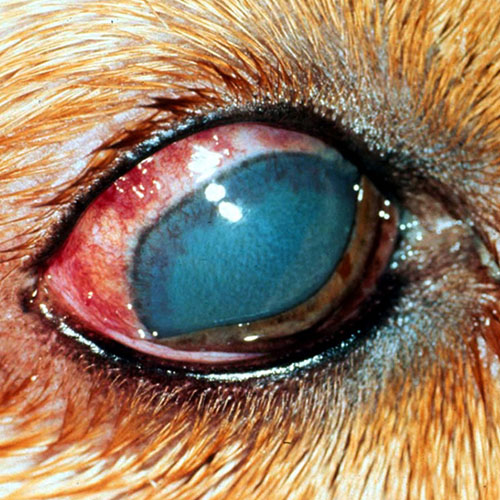
SYMPTOMS
Clinical signs of glaucoma include redness in the white part of the eye (bloodshot eye), a cloudy or hazy cornea, and a dilated pupil. As glaucoma is a throbbing pain, it can be difficult to assess whether or not your pet is painful. Some pets will squint or hold their eye shut. This is a clear indication of pain in our companion animals. Third eyelid elevation can also be a sign of pain. Blindness is yet another symptom of glaucoma though it can be difficult to recognize when only one eye is affected.
With chronic or long-standing glaucoma, the eye itself can stretch and become enlarged. Glaucoma tends to have an insidious onset and often times the first eye is already permanently blind when evaluated by the ophthalmologist. Much attention will therefore be focused on saving the remaining eye.
CAUSE
Glaucoma is either primary or secondary. Primary glaucoma occurs without evidence of previous ocular disease and is thought to have an inherited basis. Certain breeds are known to be at high risk for glaucoma. These include, but are not limited to: Cocker Spaniels, Bassett Hounds, artic breeds such as Siberian Huskies, and Chow Chows.
Secondary glaucoma is caused by some form of ocular disease that decreases fluid egress from the eye. These conditions can include inflammation, lens instability, trauma to the eye, bleeding inside the eye, as well as cancerous processes.
Your veterinary ophthalmologist will perform a complete ocular examination in order to determine if you pet is affected with primary or secondary glaucoma. The ophthalmologist may perform gonioscopy by temporarily placing a contact lens on your pet’s eye. This allows the ophthalmologist to evaluate the drainage angle of the eye. This can be important in determining the risk for glaucoma in the unaffected eye.
DIAGNOSIS
History, clinical signs, tonometry (pressure check), and gonioscopy are used in the diagnosis of glaucoma.
Tonometry is the measurement of intraocular pressure. This gives the ophthalmologist an objective measurement which can be tracked overtime to assess response to treatment. Applanation and rebound tonometry are the standard of care today. The surface of the eye will be numb so that your pet is comfortable while the pressure is measured. This test is noninvasive and takes only a few seconds.
TREATMENT
The treatment of glaucoma must be instituted as soon as possible. If pressures remain elevated for even 24 hours, irreversible damage can occur to the retina and optic nerve. Unfortunately this leads to permanent blindness in the affected eye.
Our goals in the treatment of glaucoma are two-fold: to maintain vision and to maintain comfort.
Aggressive medical therapy will be instituted once glaucoma is diagnosed. At times, mannitol is administered intravenously to rapidly decrease intraocular pressure. If the pressure remains high, a small amount of fluid can be removed from the eye. Once the pressure is within a normal range, topical medications such as dorzolamide, latanoprost, timolol, and demecarium bromide will be prescribed to maintain a normal intraocular pressure. These medications either decrease fluid production inside the eye or improve outflow of fluid from the eye.
Each animal is unique in how they respond to medical management. Certainly medical management can fail, especially in the long-term control of glaucoma.
If medical management fails, several surgical options are available. The type of surgery is based whether the eye has maintained vision. For visual eyes, laser surgery is available to decrease fluid production by killing the cells that produce fluid. Gonio implants are also available. These increase fluid outflow by placing a small tube into the eye which allows fluid to leave the eye through a valve that opens in response to elevated pressures. These procedures may be combined. Complications do exist with these procedures. Complications include inflammation, intraocular hemorrhage, infection, migration or clogging of the tube, and phthisis (shrinking of the eye).
If the eye is blind, surgeries of comfort are considered. An eye that is blind and non-painful does not require surgery. Often times, however, glaucomatous eyes are blind and very painful. Signs of pain can be subtle in animals and may go unrecognized. Often pets will sleep more, play less, have a decreased appetite or water consumption, and become less interactive. These symptoms can develop slowly overtime and therefore be difficult to recognize. We know from experience that animals are affected by the pain of glaucoma. When surgeries of comfort are performed, clients often report that their pet’s quality of life has greatly improved. Surgeries of comfort include eye removal or a prosthetic/false eye.
PROGNOSIS
Despite advances in medical and surgical management of glaucoma, the condition typically remains progressive and is the leading cause for blindness in companion animals. Medical and surgical management often fail to maintain vision and comfort long-term.
Prognosis is based on the condition of the eye at initial evaluation, response to medical and surgical therapy, consistent medication administration, as well as consistent follow-up examinations and pressure checks.