 Located in the SAVS Facility
CATARACTS
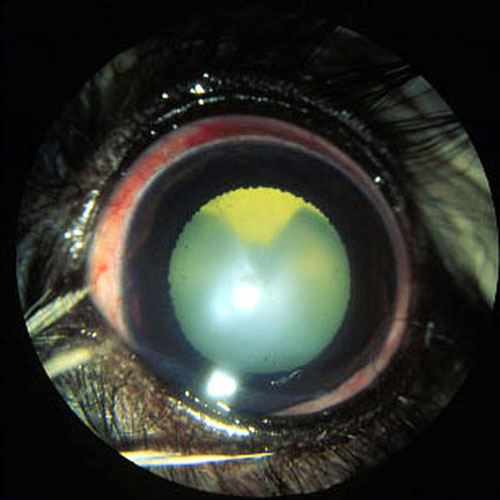
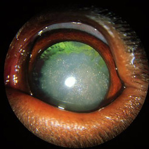
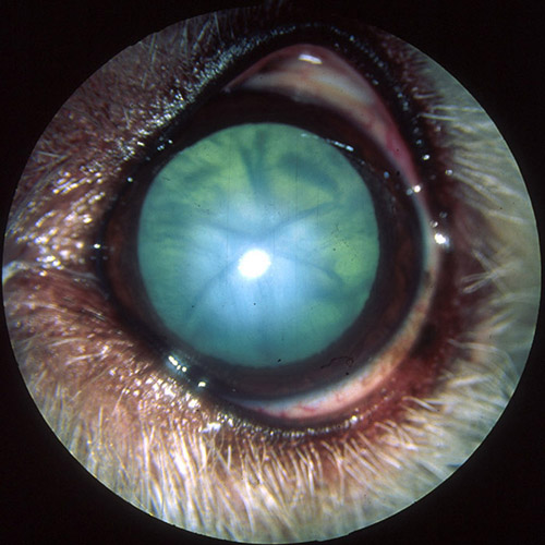
A cataract is defined as any opacity or cloudiness to the lens. The lens is a structure inside that eye that focuses light on the retina. Cataracts scatter or block light and prevent light from reaching the retina.
The lens is made up of protein surrounded by a very thin, clear elastic shell or capsule. The lens is naturally clear because it is highly organized. If this highly organized structure is disrupted, a cataract occurs. Since a cataract is a physical change in the protein structure of the lens, there are no drops (human or veterinary) that can dissolve a cataract or halt its progression.
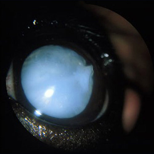
Cataracts can be in seen one eye or both eyes. Cataracts come in many shapes and sizes from small pinpoint dots to a diffuse white color that fills the entire lens. If the cataract fills the entire lens, it can lead to blindness. It is very difficult to predict whether a small cataract will progress and if so how fast it will progress. Each cataract is unique.
CAUSES OF CATARACTS
A common and important cause for cataracts is diabetes mellitus. All dogs with mature or complete cataracts should have blood work performed to test for diabetes.
Cataracts can also be inherited. Even if both parents are free of cataracts, they can still produce offspring that develop cataracts. Many times if no other ocular abnormalities are noted on exam and no clear cause is found for the cataracts, they are assumed to be inherited.
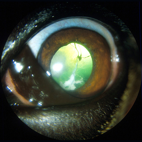
Certainly trauma or inflammation inside the eye can precipitate cataract formation. In fact, inflammation inside the eye is the most common cause of cataract formation in cats.
HOW DO YOU TREAT CATARACTS?
Treatment recommended for the cataract will often depend on the size and position of the cataract. Treatment is also based on how the presence of the cataract is affecting your pet’s quality of life.
Topical anti-inflammatory eye drops are often recommended to prevent unwanted side effects from cataracts such as inflammation inside the eye and glaucoma (high pressure inside the eye).
Eye drops alone will not resolve a cataract or slow its progression. They are simply meant to keep your pet comfortable long-term.
If cataracts are affecting vision and the goal is to restore vision in order to improve quality of life, cataract surgery is necessary. In both the human and veterinary medical fields, surgery is the only known treatment shown to restore vision. In fact, cataract surgery in humans is one of the major Medicare expenses each year.
MY PET HAS A CATARACT – WHAT SHOULD I DO?
If you suspect your pet is developing a cataract, the first step would be to schedule an initial examination with the ophthalmologists at South Texas Veterinary Ophthalmology. The ophthalmologist will perform a thorough examination from the front to the back of the eye. After the exam, they will be able to describe the cataract and any other ocular findings to you in detail. Cataract surgery is not necessary in all cases.
The sooner your pet is examined the better. Left alone, cataracts can lead to vision threatening inflammation, glaucoma, and retinal detachments. Surgery performed on cataracts which have just recently developed carries the highest success rate. We do not need to wait for cataracts to mature.
If cataract surgery is recommended, your veterinary ophthalmologist will discuss the procedure and recovery with you in detail.
Prior to surgery, several diagnostics tests are required to ensure your pet is a good candidate for cataract surgery. These include, but are not limited to, an electroretinogram (to ensure the retina is functioning), an ocular ultrasound (to ensure the retina is attached), gonioscopy (to evaluate your pet’s risk for glaucoma), and a full blood work panel (to help us assess your pet’s overall health).
WHAT’S INVOLVED IN THE ACTUAL SURGERY?
Cataract surgery in animals is performed with the same equipment used by human surgeons. Your pet will be fully anesthetized for the procedure. Patient vitals such as heart rate, EKG, blood pressure, temperature, CO2, oxygenation status, and respiratory rate are continuously monitored throughout the procedure.
A very small incision is made in the cornea to allow the surgeon access to the lens. A small circular area of lens capsule is also removed. Ultrasound power is then employed to break up the abnormal lens and remove it from the eye. Once this is complete, only the clear elastic bag (capsule) of the lens remains. Our goal is to then implant a new acrylic artificial lens inside this capsule to bring your pet’s vision back to focus. As the entire cataract has been removed, cataracts cannot regrow.
Stitches or sutures are placed in the small corneal incision to help it to heal.
Patients are monitored closely as they recover from anesthesia. We will also begin to closely monitor intraocular pressure and administer appropriate post-operative eye medications.
Your pet will stay over night for continued care and be sent home with you the next day.
Once you bring your pet home, you can expect a regimen of oral medications and topical eye drops to help the eye heal and protect against infection. The details of this regimen will be thoroughly explained when your pet is discharged from the hospital. It is imperative that these medications are administered. The medications will be tapered or decreased gradually throughout the recovery period.
Full healing from cataract surgery is approximately 6 – 8 weeks. Your pet will be able to see immediately after surgery. The healing time takes into account inflammation that requires treatment after surgery as well as time for the new lens to scar in place and the corneal incision to heal. Your pet will be re-evaluated by the ophthalmologist several times during this 6 – 8 week recovery period.
DOES CATARACT SURGERY HAVE COMPLICATIONS?
Yes, as with any surgery, there are complications that can occur. The overall success rate with cataract surgery is 90%.
10% of patients can experience one or more of the following complications:
- Retinal detachment
- Glaucoma
- Chronic inflammation
- Breakdown or infection of the incision site
- Corneal ulcerations
- Displacement of the intraocular lens implant
- Infection inside the eye
Several of these complications can lead to pain and/or blindness. Our veterinary ophthalmologists are well aware of the potential complications with cataract surgery and will monitor your pet closely.
WHAT DO PATIENTS SEE AFTER THE SURGERY?
Cataract surgery is a very rewarding procedure. Often times, pets wake up from surgery and immediately start looking around the room. Most are able to navigate immediately. The vision can become a little foggy for the first 2 weeks due to inflammation inside the eye.
There are times when an artificial lens cannot be placed. With the cataract removed, the patient will still have excellent vision (the lens is simply used for focusing up close). These patients will be able to see very well far away. Near sighted vision may be a little unfocused (much like a human that needs reading glasses). As our companion animals do not read or drive the difference in near and far vision is not often noticed. The artificial lens implant is icing on the cake for cataract surgery – the most important aspect of the surgery is to remove the abnormal lens to clear the line of sight.