 Located in the SAVS Facility
PROLAPSED GLAND OF THE THIRD EYELID (CHERRY EYE)
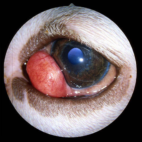
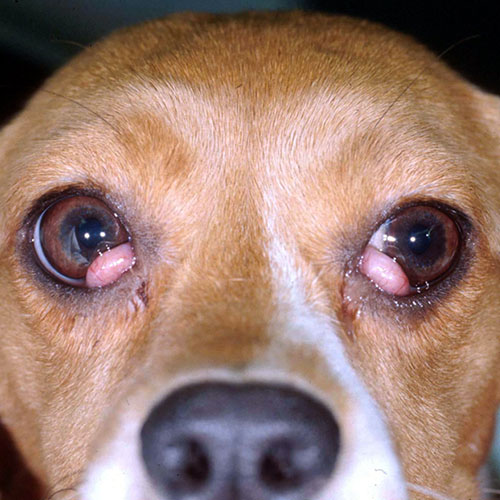
Prolapsed gland of the third eyelid is often referred to as “Cherry Eye.” This condition is more common in dogs than cats. It typically affects dogs that are less than 2 years of age but has been observed in older pets. It can affect one or both eyes.
The prolapse occurs due to a weakness of the tissues that hold the gland in place. The gland typically lives out of sight at the base of the third eyelid, deep in the inside corner of the eye. When the tissues are weak the gland is allowed to rise and become visible at the leading edge of the third eyelid. At that time, a smooth red mass will be seen. This is not a painful condition; however, overtime the gland can become inflamed due to exposure and dryness. With increased inflammation the gland can even bleed.
The gland of the third eyelid is important as it is responsible for 30% of the aqueous tear production. Allowing the gland to remain prolapsed or removing the gland will put your pet at increased risk for conjunctivitis and dry eye. Dry eye leads to corneal ulcers, conjunctivitis, ocular infections, patient discomfort, corneal vascularization, corneal scarring, and corneal mineralization.
TREATMENT
The gold standard of treatment is to surgically reposition the gland. Removal of the gland is not recommended. Surgery has a 90% success rate. Based on ophthalmic examination and the age of your pet, the doctor may recommend preventatively treating the unaffected eye.
Healing time from surgery is approximately 2 weeks. An e-collar or cone will be required during this time to protect the surgery site. Activity should be kept to a minimum. Oral and topical medications will be prescribed to decrease inflammation and prevent infection. Recovery from this procedure is typically uneventful and swelling is minimal.
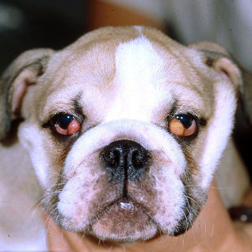
As with any surgery, complications do exist. The most common complication for this surgery is recurrence of the gland prolapse. Breeds such as English Bulldogs and Mastiff breeds are at high risk for recurrence. Typically this necessitates a second surgery. Other rare complications include conjunctivitis, inflammation, breakdown of the suture, infection, corneal ulcers, or corneal scarring.