 Located in the SAVS Facility
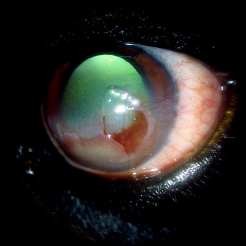
INDOLENT CORNEAL ULCER
(Non-healing ulcer, Boxer ulcer, spontaneous chronic corneal epithelial defect)
The cornea is the clear windshield to the eye. Together with the sclera (the white part of the eye), the cornea makes the outside wall or shell of the eye. The cornea must be clear to allow light to enter the eye for vision. This is achieved by its highly organized layers, lack of blood vessels, and a special layer that pumps water out of the cornea to maintain its dehydrated state.
The cornea is made up of many layers. The surface layer of cells is called the epithelium. The middle layers are referred to as the stroma of the cornea and the very inside layer is called the endothelium.
Superficial Ulcers
The term corneal ulcer refers to a break or wound in the cornea. If this wound only involves loss of the first layer of cells, the epithelium, it is called a superficial ulcer.
Despite being surface wounds, superficial ulcers can be very painful. Loss of the first layer of cells allows the corneal nerves to be exposed which are the cause for the pain and irritation felt by the patient. The cornea heals very quickly and with appropriate therapy, superficial ulcers should heal within 48 hours.
Indolent Ulcers
Indolent corneal ulcers are superficial ulcers that do not heal in the appropriate amount of time. They are a specific type of ulcer and are typically age related (occur in middle aged and older pets). As dogs and cats age, the adherent layer between the epithelium and underlying stroma degenerates. Thus, the epithelium floats on the surface of the eye and is easily peeled back or removed. Indolent ulcers in dogs as well as humans are spontaneous and do not require an inciting cause. These ulcers will follow a course of appearing to improve and then rapidly worsening. As the epithelium attempts to grow and heal the ulcer, large flaps of tissue are formed. These cover the corneal nerves and make the patient more comfortable temporarily. Without the “glue” to hold them in place, however, these new cells are easily removed from the cornea and symptoms return. Therefore, indolent ulcers remain present for months without appropriate treatment.
SYMPTOMS
Patients with indolent ulcers typically exhibit one or more of the following clinical signs or symptoms:
- Squinting or holding the eye shut
- Redness in the white part of the eye
- Discharge from the eye. This discharge can be clear, white, yellow, or green
- Redness or cloudiness on the surface of the eye
- Rubbing at the eye
These symptoms may fluctuate showing signs of improvement followed by deterioration.
DIAGNOSIS
The primary diagnostic test for an indolent corneal ulcer is fluorescein stain. The stain will bind to the injured portion of the cornea. A slit lamp biomicroscope is then used to evaluate the cornea under a high degree of magnification. The extent and depth of the ulcer is noted. The flaps of tissue discussed above can be observed by the ophthalmologist under the slit lamp microscope.
Further diagnostic testing such as corneal cytology and corneal culture may be recommended.
TREATMENT
The primary focus of treatment for indolent corneal ulcers is restoring the adherent layer or glue between the epithelium and stroma. Medical management alone is often unsuccessful.
Surgical therapy in the form a corneal debridement is the first step in treating indolent ulcers. This allows the affected cells to be removed from the cornea. Once this is complete, micro abrasions are created in the corneal stroma. This allows new cells to have a place to attach as they grow across the cornea. Combined, these procedures carry a 90% success rate of healing the ulcer in 2 weeks. In some cases, the procedures need to be repeated a second time for complete healing.
Often, a bandage contact lens is placed over the cornea once the procedures are complete. This will protect the ulcer as it heals and make your pet more comfortable during the healing process.
Topical as well as oral medications will be required after the procedure to protect against infection as well as control pain and inflammation. Once the ulcer is fully healed, topical antiinflammatory drops may be recommended to decrease any scar tissue that may have formed.
PROGNOSIS
With the appropriate therapy, the prognosis for vision as well as comfort is very good. It is very rare for an indolent ulcer to recur after appropriate treatment.
Once one eye is affected, the fellow eye is at risk for a similar indolent ulcer in the future. There are currently no known medications or treatments to prevent the fellow eye from developing an indolent ulcer.