 Located in the SAVS Facility
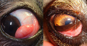
Dogs and cats have a third eyelid? They sure do! The third eyelid, also known as the nictitating membrane, is a triangular, mobile, protective, and glandular structure lying between the cornea and the lower eyelid. The majority of the third eyelid is hidden at the inside corner of our pet’s eyes.
The third eyelid has several functions such as distribution of the tear film, protection of the cornea, and production a portion of the tear film. The gland of the third eyelid produces between 30% and 50% of the normal tear film in dogs.
So what can happen to the third eyelid? The most noticeable (and at times surprising) condition that can occur is a protrusion of the gland of third eyelid known colloquially as “cherry eye”. When this occurs, a red round mass is noted at the leading edge of the third eyelid (see picture 1). The gland should be surgically replaced to retain its function and to prevent the exposed gland from becoming dry, inflamed, or secondarily infected. Many years ago, veterinarians recommended removing the glands. It is now known that prolapsed glands should be surgically replaced and never removed. Studies confirm that removing a gland will put the patient at high risk for severe dry eye and chronic inflammation of the cornea and conjunctiva (white part of the eye).
Elevation of third eyelid can also occur. This often creates the illusion that the pet’s eye is rolling back into the head. The eye itself is in fact in a normal position but the third eyelid begins to cover it (see picture 2). There are many causes for an elevated third eyelid: Horner’s syndrome (a neurologic disease usually seen in older dogs and cats), a mass or abscess behind the eye, a small globe, active retraction of the globe in painful eye conditions or due to loss of orbital contents as in dehydration, emaciation, or scarring. If you notice this symptom in your pet, take your pet to a veterinarian for an examination.
At times, foreign bodies become lodged between the eye and the third eyelid causing pain. These can be anything from a blade of grass, to cactus thorns and grass awns. Injuries to the third eyelid occur as a result of fights, motor vehicle accidents, and foreign body penetration. Small tears may not require stitches. Larger lacerations may benefit from careful placement of stitches by a veterinarian or veterinary ophthalmologist.
As you can see, the third eyelid is a useful and important structure! The only reasons for its removal are severe irreparable trauma and cancer of the third eyelid. Any other conditions can be successfully treated by your veterinarian or your veterinary ophthalmologist.