 Located in the SAVS Facility
CORNEAL ULCERS
The cornea is the clear windshield to the eye. Together with the sclera (the white part of the eye), the cornea makes the outside wall or shell of the eye. The cornea must be clear to allow light to enter the eye for vision. This is achieved by its highly organized layers, lack of blood vessels, and a special layer that pumps water out of the cornea to maintain its dehydrated state.
The cornea is made up of many layers. The surface layer of cells is called the epithelium. The middle layers are referred to as the stroma of the cornea and the very inside layer is called the endothelium. Based on these layers, there are several types of corneal ulcers: superficial, stromal, and descemetoceles.
Superficial Ulcers
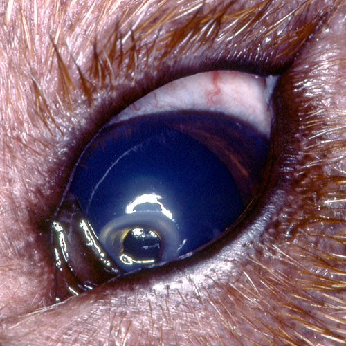
The term corneal ulcer refers to a break or wound in the cornea. If this wound only involves loss of the first layer of cells, the epithelium, it is called a superficial ulcer.
Despite being surface wounds, superficial ulcers can be very painful. Loss of the first layer of cells allows the corneal nerves to be exposed which are the cause for the pain and irritation felt by the patient. Luckily, the cornea heals very quickly and with appropriate therapy, superficial ulcers should heal within 48 hours.
Stromal or Complicated Ulcers
Ulcers that do not heal within the first few days of treatment, become infected, or involve the deeper layers of the cornea are referred to as complicated ulcers.
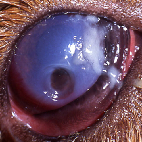
Complicated ulcers can occur for a variety of reasons. First, repeated trauma to the surface of the eye by a foreign body or extraneous hair will delay healing indefinitely. Secondly, other ocular conditions such as dry eye, glaucoma, and inflammation can also lead to serious corneal ulcers. Lastly, ulcers can worsen due to infection. There are naturally bacteria around the eye. If there is a wound on the eye, these bacteria take the opportunity to colonize the cornea and can lead to rapid corneal degradation. Sometimes, infected ulcers are referred to as “melting” ulcers because the cornea becomes very soft due to enzymatic degradation of the cornea.
Descemetoceles
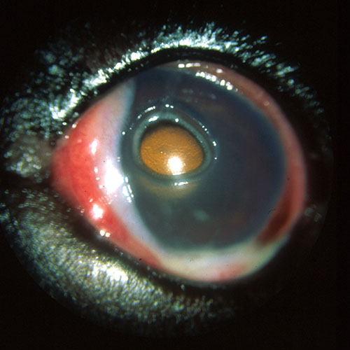
The term descemetoceles refers to an ulcer so deep that only one layer of corneal cells remain. Descemetoceles are impending perforations as this last layer of cornea is extremely fragile.
SYMPTOMS
DIAGNOSIS
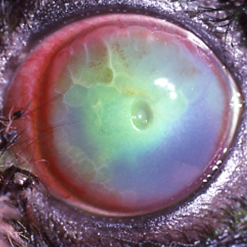
The primary diagnostic test for a corneal ulcer is fluorescein stain. The stain will bind to the injured portion of the cornea. A slit lamp biomicroscope is then used to evaluate the cornea under a high degree of magnification. The extent and depth of the ulcer is noted. The slit lamp is also used to look for the cause of the ulcer (ex: foreign body, extra hairs, or a poor quality tear film).
Depending on the type of ulcer diagnosed, further diagnostic testing such as corneal cytology and corneal culture may be recommended.
TREATMENT
Medical as well as surgical management may be recommended for your pet’s ulceration.
Medical management involves the use of antibiotics, anti-inflammatories, pain medicine, drops to dilate the pupil, and at times a blood product called serum. These will aid in healing the ulceration as well as making your pet more comfortable. Bandage contact lenses can also be used to protect the ulcer as it heals as well as cover the exposed corneal nerves.
If an ulcer involves 50% or more of the corneal thickness, surgery may be recommended to avoid corneal perforation and loss of vision. Surgical techniques include conjunctival grafts, corneoconjunctival grafts, and use of other biological graft material to stabilize the cornea. These grafts become incorporated into the patient’s cornea.
PROGNOSIS
If treatment is sought early, the vast majority of ulcers will heal and vision will be saved.
Corneal surgery typically carries a 90% success rate. As with any surgical procedure, complications (including potential anesthetic risks) exist. Potential complications include conjunctivitis, wound dehiscence, graft rejections, infections at the surgical site or inside the eye, corneal scarring, corneal vascularization, corneal mineralization, inflammation inside the eye, glaucoma, retinal detachment, bleeding inside the eye, and excessive tearing.
Our goal with medical or surgical treatment of corneal ulcers is to give your pet the best vision possible and to control any pain or discomfort they may be experiencing.